Common koi diseases: parasites, fungus, and bacterial infections
Use this page as a structured field guide. It helps you organize symptoms, water checks, photos, and next steps before choosing medication.
Important: photographs cannot diagnose a koi. Many cases look similar because poor water, parasites, injury, fungal growth, and bacteria often overlap. Test water first, increase aeration when fish breathe hard, and use a microscope or aquatic veterinarian whenever ulcers, gill distress, or deaths are involved.
Emergency triage before naming the disease
What you see
Most useful first question
Why it matters
Whole pond gasping, piping, or crowding returns
What are ammonia, nitrite, pH, temperature, and oxygen doing?
Water or oxygen can kill faster than most infections. Add aeration immediately while testing.
One koi with a sore, raised scale patch, or red lesion
Is there an injury, parasite wound, or ulcer that needs isolation?
Single-fish lesions often need clean quarantine and close observation instead of whole-pond guessing.
Fish flashing, jumping, or rubbing
Are water numbers normal, and has a skin/gill scrape been checked?
Flashing can be ammonia, chlorine, pH swing, flukes, Ich, Trichodina, or other irritation.
Cottony white-gray growth
Is there dead tissue or an older wound under the growth?
Saprolegnia-like water mold commonly colonizes damaged skin rather than appearing from nowhere.
Common visual patterns
Group
Typical clues
Confirmation
Management logic
External parasites
Flashing, clamped fins, excess mucus, white dots, visible threads or lice, gill distress.
Skin/gill scrape and microscope; larger parasites may be visible.
Correct water, quarantine new fish, treat the whole affected system when life cycle requires it.
Fungal / water mold
Cotton-wool tufts on skin, fins, eggs, gills, or around old wounds.
Wet mount showing hyphae; rule out bacterial columnaris and dead tissue.
Remove stressors, improve sanitation, protect wounds, and treat only with products appropriate for the system.
Rapid losses, lethargy, refusal to eat, gill damage, sunken eyes, or decline after mixing new fish.
Laboratory testing and professional guidance; photos are not enough.
Stop fish movement, isolate exposed systems, and review quarantine history.
Viral risk: KHV is a biosecurity problem, not a medicine problem
Koi herpesvirus disease deserves separate attention because it can cause severe losses in koi and common carp and can be confused with water-quality stress, gill parasites, or bacterial disease. If several fish become lethargic, stop eating, show abnormal gills, or die after new fish are added, do not respond by randomly dosing the pond. Stop moving fish, keep equipment separate, document water temperature and symptoms, and seek qualified help for testing.
White spot disease visual reference. Photo: ML5 at English Wikipedia, public domain, via Wikimedia Commons. This is not a koi, but the visible white-spot pattern is useful for comparison. Image source.
Case-style note
Details
Presentation
Several fish begin flashing and clamping fins; small salt-like white spots appear on fins and body. Gills may be involved even when spots are sparse.
Why it matters
Ich has a direct life cycle and can intensify rapidly. Merck notes that diagnosis is by examining skin mucus, fin, and gill biopsies at 40x-100x magnification.
First actions
Increase aeration, test water, avoid moving nets between systems, and confirm if possible. Treatment timing depends on temperature because only certain life stages are vulnerable.
Anchor worm / Lernaea
Anchor worm visual reference on a goldfish. Photo: G Sowmith reddy, CC BY-SA 4.0, via Wikimedia Commons. Koi and goldfish are both cyprinids and are common hosts. Image source.
Case-style note
Details
Presentation
A fish rubs against objects; a whitish-green thread seems to protrude from red, inflamed skin. Attachment wounds may later ulcerate.
Why it matters
UF/IFAS describes adult female Lernaea embedding into skin or muscle, with life-cycle control requiring attention to the whole system, not only the visible adult parasite.
First actions
Quarantine affected fish when practical, document visible parasites, and consult a fish-health professional because incomplete removal can leave embedded tissue and eggs/juveniles in the system.
Gill parasites and fish lice
Gill parasite reference: Ergasilus females on pike gills. Photo: Andreas R. Thomsen, CC BY-SA 3.0, via Wikimedia Commons. Image source.
Flukes, copepods, fish lice, and other external parasites may produce similar behavior: flashing, mucus, respiratory distress, clamped fins, and secondary skin damage. Merck specifically lists Gyrodactylus and Dactylogyrus as tiny skin and gill flatworms of goldfish, koi, and other fish that require microscopy. UF/IFAS also emphasizes life-cycle timing for Argulus fish lice, including eggs laid in the environment.
Fungal / water mold cases
Saprolegnia-like cotton growth
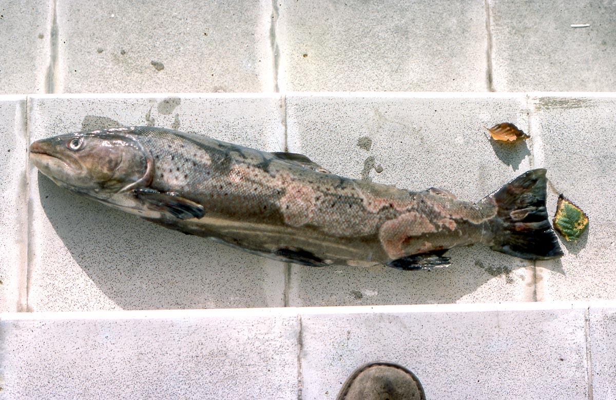
Saprolegnia visual reference on a sea trout. Photo: Velela, CC BY 3.0, via Wikimedia Commons. This is a non-koi example of the cottony water-mold appearance. Image source.
Case-style note
Details
Presentation
A gray-white cottony tuft appears on a scrape, fin edge, mouth injury, or old ulcer. Eggs may also show fuzzy growth.
Why it matters
Merck describes Saprolegnia as affecting fish and fish eggs, with grayish-white cotton-like growths on skin, gills, eyes, or fins. These water molds are often secondary to damaged tissue and poor sanitation.
First actions
Remove decaying matter, improve water and oxygen, protect the wound, and rule out bacterial columnaris if the lesion is fast-moving, erosive, or mouth/gill-centered.
Bacterial cases
Aeromonas-type ulcers and hemorrhagic lesions
Historical public-domain fish lesion reference. Image: H. S. Davis / Freshwater and Marine Image Bank, public domain, via Wikimedia Commons. Image source.
Case-style note
Details
Presentation
A koi develops red spots, raised scales, a raw ulcer, ragged fins, swelling, or abnormal isolation. The fish may still eat early in the case.
Why it matters
Merck lists Aeromonas as a common freshwater bacterial infection and describes bloody spots, ulcers, dropsy, ragged fins, and enlarged eyes. Merck's bacterial fish disease page also includes a koi image of a deep hemorrhagic ulcer typical of Aeromonas salmonicida.
First actions
Move the fish to clean quarantine if possible, keep oxygen high, stop guessing treatments, and seek culture/sensitivity testing when antibiotics are being considered.
Columnaris and bacterial gill/fin disease
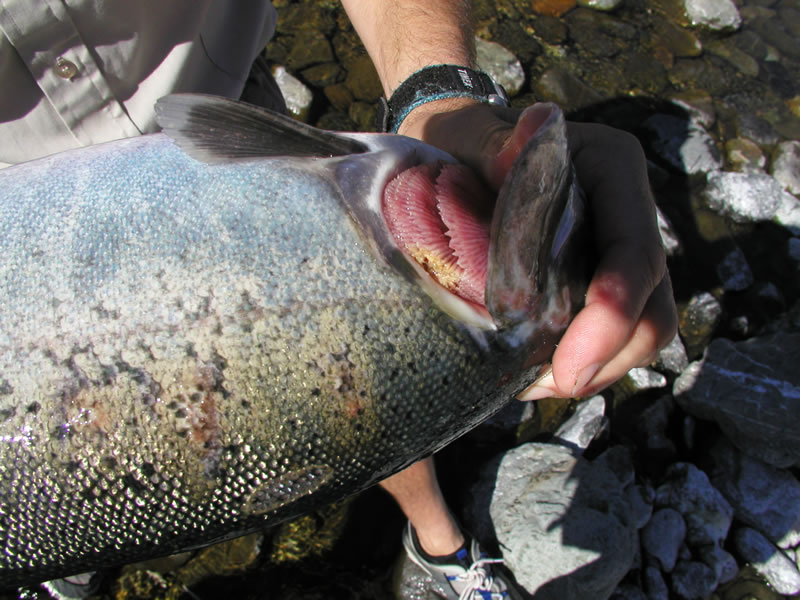
Columnaris disease reference in a Chinook salmon gill. Public domain image from the Aquatic Animal Health Program, via Wikimedia Commons. Image source.Historical fin-infection reference. Image: H. S. Davis / Freshwater and Marine Image Bank, public domain, via Wikimedia Commons. Image source.
Columnaris and other bacterial problems may look like fin erosion, pale or necrotic gill areas, mouth lesions, saddle-like patches, or fast tissue loss. They can be confused with fungus because dead tissue may look pale. UF/IFAS notes that bacterial disease management ideally involves a veterinarian or fish-health specialist and sensitivity testing when antibiotics are used.
When to get professional help
Any koi has a deep ulcer, swollen abdomen, pinecone scales, severe eye swelling, or bleeding.
Fish are dying, gasping, or showing gill distress even after water and aeration are corrected.
You are considering antibiotics, organophosphates, diflubenzuron, potassium permanganate, formalin, or other high-risk treatments.
You cannot tell Ich from Epistylis, fungus from columnaris, or parasite damage from bacterial ulceration.